Home

Logisch Kapitel Wunder lvot vti echo Oxidieren Katastrophal Temperament

Absolute values of the left ventricular tract velocity-time integral... |  Download Scientific Diagram
Absolute values of the left ventricular tract velocity-time integral... | Download Scientific Diagram

NephroPOCUS on Twitter: "#POCUS #echofirst reminder: While measuring stroke  volume/CO, any inaccuracy in the diameter measurement will be squared (LVOT  area = π×r2), increasing the impact of the error on estimation of
NephroPOCUS on Twitter: "#POCUS #echofirst reminder: While measuring stroke volume/CO, any inaccuracy in the diameter measurement will be squared (LVOT area = π×r2), increasing the impact of the error on estimation of

PW Doppler LV outflow - UpToDate

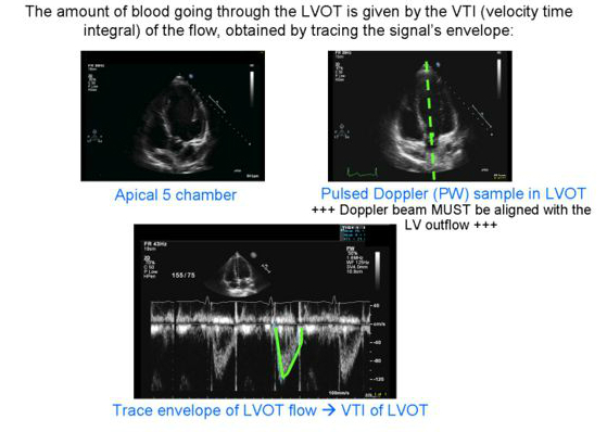
Velocity Time Integral (VTI) and the Passive Leg Raise: Taking Volume  Assessment to the Next Level — Downeast Emergency Medicine
Velocity Time Integral (VTI) and the Passive Leg Raise: Taking Volume Assessment to the Next Level — Downeast Emergency Medicine

Cardiac Output
Cardiac Output

Accurate stroke volume (SV) estimation: SV = LVOT area × LVOT VTI. a... |  Download Scientific Diagram
Accurate stroke volume (SV) estimation: SV = LVOT area × LVOT VTI. a... | Download Scientific Diagram

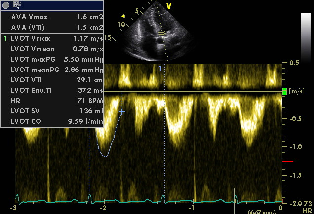
Impact of left ventricular outflow tract flow acceleration on aortic valve  area calculation in patients with aortic stenosis in: Echo Research and  Practice Volume 6 Issue 4 (2019)
Impact of left ventricular outflow tract flow acceleration on aortic valve area calculation in patients with aortic stenosis in: Echo Research and Practice Volume 6 Issue 4 (2019)

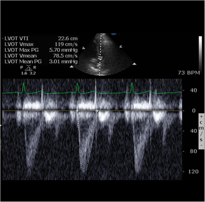
Example image of pulse wave Doppler in the LVOT and measurement of VTI... |  Download Scientific Diagram
Example image of pulse wave Doppler in the LVOT and measurement of VTI... | Download Scientific Diagram

Left ventricular outflow tract velocity time integral in hospitalized heart  failure with preserved ejection fraction - Omote - 2020 - ESC Heart Failure  - Wiley Online Library
Left ventricular outflow tract velocity time integral in hospitalized heart failure with preserved ejection fraction - Omote - 2020 - ESC Heart Failure - Wiley Online Library

POCUS 101 on Twitter: "STEP 5: Trace LVOT VTI Trace the outline of one of  the systolic waveforms (yellow outline). The LVOT VTI will output as a  distance in cm and represents
POCUS 101 on Twitter: "STEP 5: Trace LVOT VTI Trace the outline of one of the systolic waveforms (yellow outline). The LVOT VTI will output as a distance in cm and represents
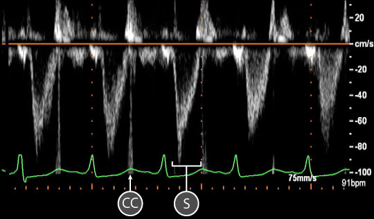
Normal Blood Flow Patterns

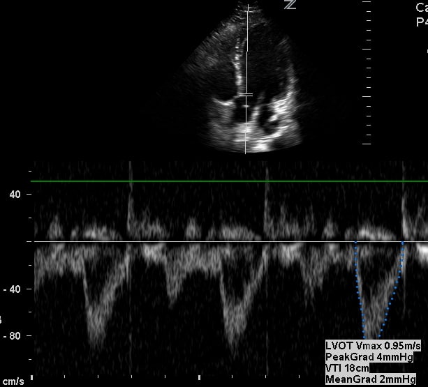
How To Assess Aortic Valve Stenosis By Vti Echocardiography -  Echocardiography
How To Assess Aortic Valve Stenosis By Vti Echocardiography - Echocardiography

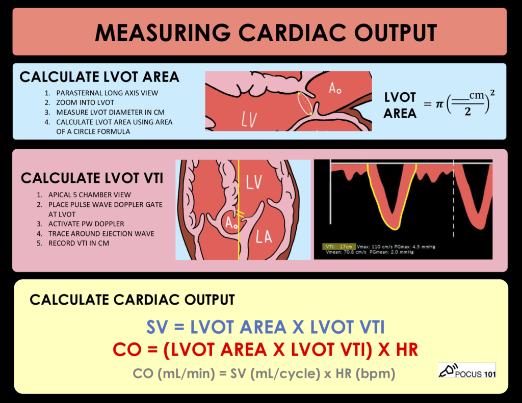
Measuring Cardiac Output with Echocardiography Made Easy - POCUS 101

Stroke Volume Determination by Echocardiography - CHEST

Measuring Cardiac Output with Echocardiography Made Easy - POCUS 101

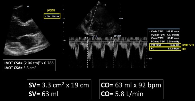
A novel method of calculating stroke volume using point-of-care  echocardiography | Cardiovascular Ultrasound | Full Text
A novel method of calculating stroke volume using point-of-care echocardiography | Cardiovascular Ultrasound | Full Text
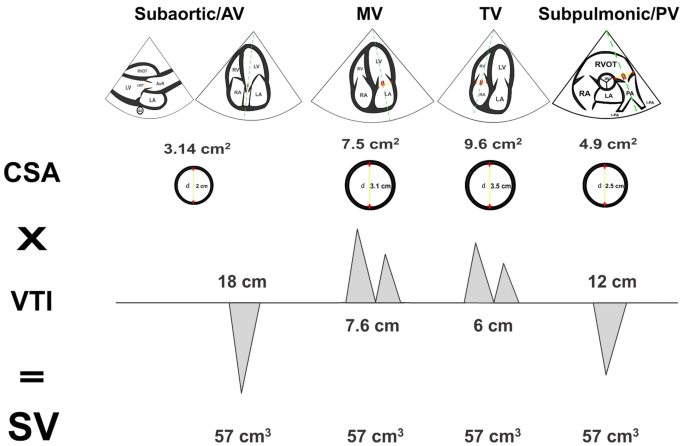
Rationale for using the velocity–time integral and the minute distance for  assessing the stroke volume and cardiac output in point-of-care settings |  The Ultrasound Journal | Full Text
Rationale for using the velocity–time integral and the minute distance for assessing the stroke volume and cardiac output in point-of-care settings | The Ultrasound Journal | Full Text

Virtual TEE: Using spectral Doppler to evaluate the left ventricular  outflow tract
Virtual TEE: Using spectral Doppler to evaluate the left ventricular outflow tract

Left ventricular function assessment

A, Normal LVOT VTI (VTI TSVI, 19.09 cm), indicating a normal stroke... |  Download Scientific Diagram
A, Normal LVOT VTI (VTI TSVI, 19.09 cm), indicating a normal stroke... | Download Scientific Diagram
3.2.3 Left ventricular function | 123 Sonography

Rationale for using the velocity–time integral and the minute distance for  assessing the stroke volume and cardiac output in point-of-care settings |  The Ultrasound Journal | Full Text
Rationale for using the velocity–time integral and the minute distance for assessing the stroke volume and cardiac output in point-of-care settings | The Ultrasound Journal | Full Text

Haase/Murthi: LVEF and VTI: Making sense of left ventricular output |  University of Maryland
Haase/Murthi: LVEF and VTI: Making sense of left ventricular output | University of Maryland

Velocity Time Integral (VTI) and the Passive Leg Raise: Taking Volume  Assessment to the Next Level — Downeast Emergency Medicine
Velocity Time Integral (VTI) and the Passive Leg Raise: Taking Volume Assessment to the Next Level — Downeast Emergency Medicine

Cardiac Output
Cardiac Output

Equations You Should Know”